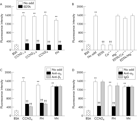
Figure 2 Connective tissue growth factor (CCN2) dependent pancreatic stellate cell (PSC) adhesion is mediated by interactions of module 4 with integrin α5β1. (A) Microtitre wells were precoated at 4°C for 16 hours with phosphate buffered saline (PBS) or 2 μg/ml CCN21–4, CCN23–4, CCN24, or fibronectin (FN) and then blocked with PBS containing 1% bovine serum albumin (BSA) for one hour. Rat activated PSC (2.5×10⁵ cells/ml) were preincubated in serum free medium for 30 minutes in vehicle buffer (no add) or EDTA (5 mM) prior to addition to individual wells at 50 μl/well. After incubation at 37°C for 20 minutes, adherent cells were washed, fixed, and stained by CyQUANT GR dye and quantified by measuring fluorescence intensity at an excitation of 485 nm and an emission of 530 nm. (B) PSC adhesion assays were performed using CCN24 following preincubation of the cells for 30 minutes with EDTA (5 mM) or with addition of Ca2+ (10 mM) or Mg2+ (10 mM) either alone or in combination. (C) PSC were preincubated with 25 μg/ml anti‐integrin α5 or anti‐integrin β1 monoclonal antibodies for 30 minutes prior to adding the cells to the wells that had been precoated with CCN24 (2 μg/ml), FN (2 μg/ml), or vitronectin (VN 4 μg/ml). (D) Microtitre wells were coated with CCN24, FN, or VN, as indicated, above prior to addition of PSC that had been preincubated at 37°C for 30 minutes with vehicle buffer (no add), 25 μg/ml monoclonal anti‐α5β1, or 25 μg/ml normal mouse IgG. Data are means (SD) of quadruplicate determinations and are representative of three experiments. **p<0.01 versus control; ††p<0.01 versus “no add” group.

An official website of the United States government
Here's how you know
Official websites use .gov
A
.gov website belongs to an official
government organization in the United States.
Secure .gov websites use HTTPS
A lock (
) or https:// means you've safely
connected to the .gov website. Share sensitive
information only on official, secure websites.
