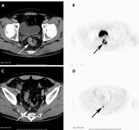
Figure 1 (A) Primary rectal tumour was exposed to the rectal wall but no extension to the pelvic side walls on computed tomography (CT) (arrow). (B) At the same level as (A), avid uptake was demonstrated on positron emission tomography (PET) (arrow). (C) A superior rectal lymph node was greater than 1 cm on CT (arrow). (D) At the same level as (C), PET showed uptake corresponded to the lymph node demonstrated on CT (arrow). This case was preoperatively diagnosed as TNM stage T3N1 and confirmed at surgery and on histopathological examination.

An official website of the United States government
Here's how you know
Official websites use .gov
A
.gov website belongs to an official
government organization in the United States.
Secure .gov websites use HTTPS
A lock (
) or https:// means you've safely
connected to the .gov website. Share sensitive
information only on official, secure websites.
