
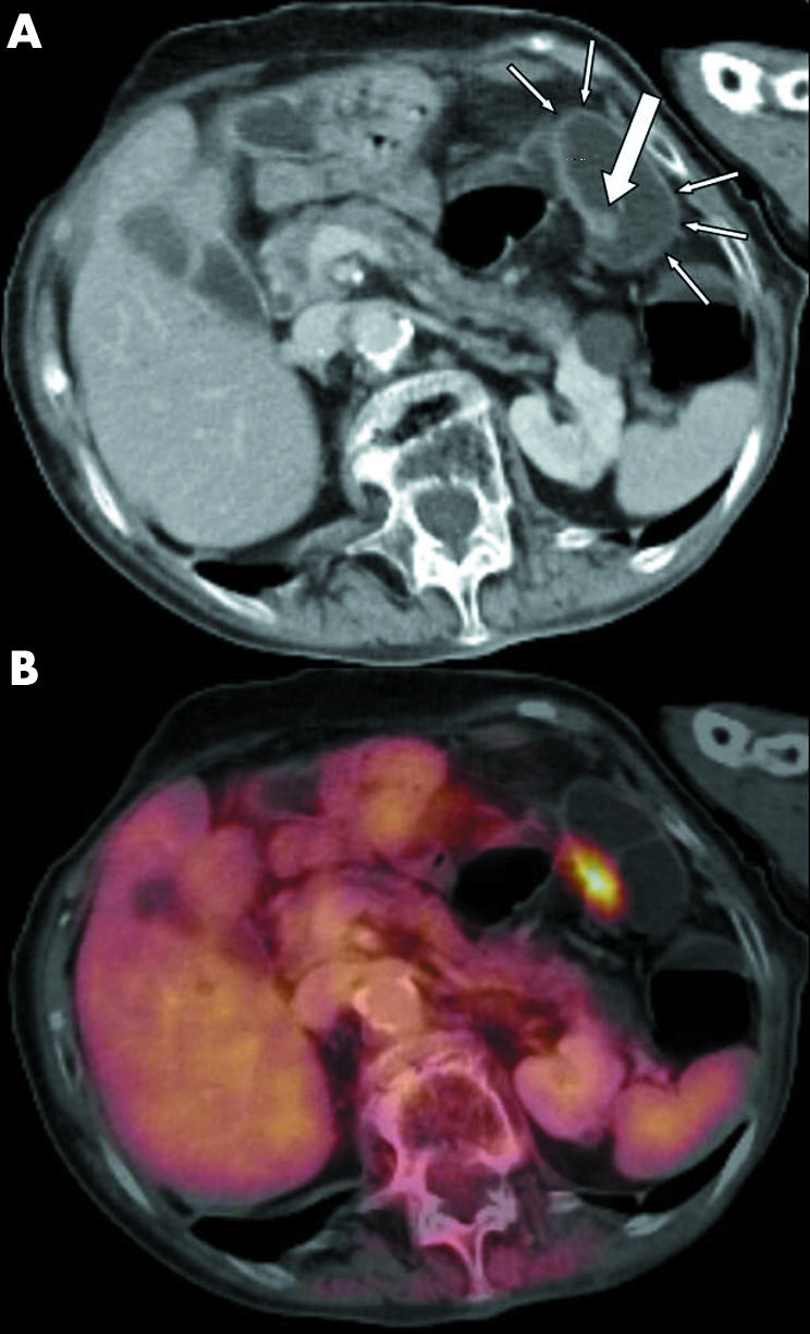
Figure 1 (A) Axial contrast enhanced computed tomography (CT) image demonstrated a tubulous polyp (diameter 9 mm) at the left colon flexure (large white arrow). The colon was well distended, rated as distension grade 3 according to the four point scale (small white arrows). (B) Corresponding axial contrast enhanced positron emission tomography/CT revealed elevated glucose metabolism within this polyp and the adjacent bowel wall, indicating a carcinoma. Verification of an adenomatous carcinoma by histopathology followed.