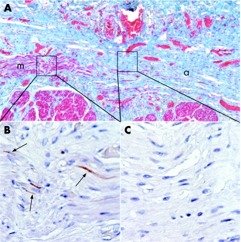
Figure 5 Interstitial cells of Cajal (ICC) are less dense in areas of smooth‐muscle atrophy than in areas of normal smooth muscle in systemic sclerosis (SSc). (A) Masson trichrome stain of the oesophagus of an SSc case with the lumenal surface towards the top of the page, showing regions of histologically normal circular smooth muscle (m), and atrophic circular smooth muscle (a). (B) C‐kit stain of a region of normal smooth muscle showing ICC (black arrows). (C) C‐kit stain of an atrophic region of smooth muscle showing no ICC. (B,C) C‐kit immunohistochemistry.

An official website of the United States government
Here's how you know
Official websites use .gov
A
.gov website belongs to an official
government organization in the United States.
Secure .gov websites use HTTPS
A lock (
) or https:// means you've safely
connected to the .gov website. Share sensitive
information only on official, secure websites.
