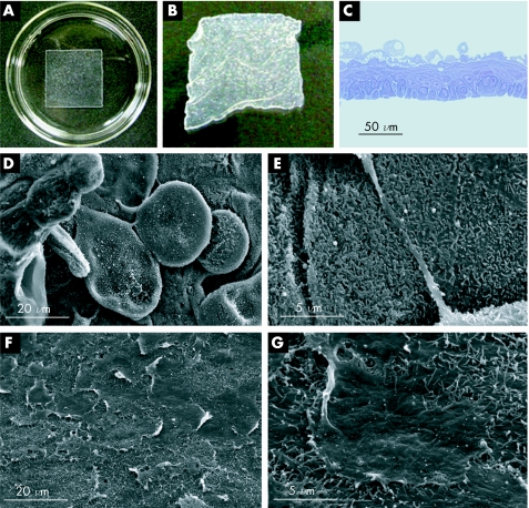
Figure 1 Transplantable oral mucosal epithelial cell sheets. (A) Under normal culture conditions at 37°C, oral mucosal epithelial cells proliferate to confluence on square‐patterned temperature‐responsive dishes. (B) By reducing the temperature to 20°C for 30 min, the cultured cells can be harvested as intact sheets, without the need for enzymatic treatments. (C) Toluidine blue staining shows that the oral mucosal epithelial cell sheets are stratified, with 3–5 cell layers. (D) Scanning electron microscopy (SEM) of the apical surface shows that the cell sheets contain flattened, polygonal superficial cells similar to the native epithelium. (E) Magnified views also show that the apical surface is covered by numerous microvilli. (F, G) SEM of the basal surface of the cell sheets shows that a basement membrane‐like deposition of extracellular matrix is maintained during low‐temperature cell sheet harvest.

An official website of the United States government
Here's how you know
Official websites use .gov
A
.gov website belongs to an official
government organization in the United States.
Secure .gov websites use HTTPS
A lock (
) or https:// means you've safely
connected to the .gov website. Share sensitive
information only on official, secure websites.
