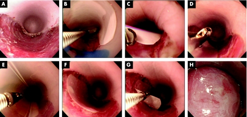
Figure 3 Endoscopic transplantation of autologous oral mucosal epithelial cell sheets. After endoscopic submucosal dissection, a flat oesophageal ulcer is created (A). The cultured oral mucosal epithelial cell sheet, attached to a white polyvinylidene difluoride (PVDF) support membrane, is then grasped by endoscopic forceps and transferred to the dissection site (B) and gently placed on the ulcer wound bed (C). After carefully withdrawing the endoscopic forceps (D), the endoscopic mucosal resection tube is used to apply gentle pressure to the PVDF support membrane and the underlying cell sheet (E). The cell sheet along with the support membrane is then left undisturbed for 10 min to allow for direct attachment to the host tissues (F). The support membrane is then easily removed (G), leaving the autologous cell sheet on the ulcer wound bed (H) (for supplementary video 1, see http://gut.bmjjournals.com/supplemental).

An official website of the United States government
Here's how you know
Official websites use .gov
A
.gov website belongs to an official
government organization in the United States.
Secure .gov websites use HTTPS
A lock (
) or https:// means you've safely
connected to the .gov website. Share sensitive
information only on official, secure websites.
