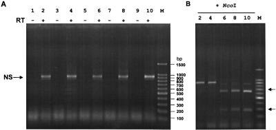
Figure 4.
Characterization of the recovered viruses by RT-PCR. (A) RNA was extracted from virus particles after two passages of the supernatant of transfected cells (see Tables 1 and 2) on MDCK cells. RT-PCR was performed with primers specific for the NS gene segment and with vRNA extracted from virions. The NS primers we used were not strain specific, thus allowing the amplification of any influenza A NS segment. The reaction products were subjected to electrophoresis on a 2% agarose gel. To ensure that the amplified DNA fragments were derived from vRNA and not from plasmid DNA carried over from transfected cells, one reaction was performed without the addition of reverse transcriptase (RT−). Lanes 1 and 2, recombinant A/Teal/HK/W312/97 (Table 1); lanes 3 and 4, M reassortant (Table 2); lanes 5 and 6, NS reassortant (Table 2); lanes 7 and 8, recombinant A/WSN/33 virus (Table 1); lanes 9 and 10, quadruple reassortant (Table 2). (B) NcoI digestion of the fragments shown in A (only the WSN-NS-cDNA has an NcoI site). The identity of the NS fragments was also verified by sequence analysis of the amplified product (not shown).

