
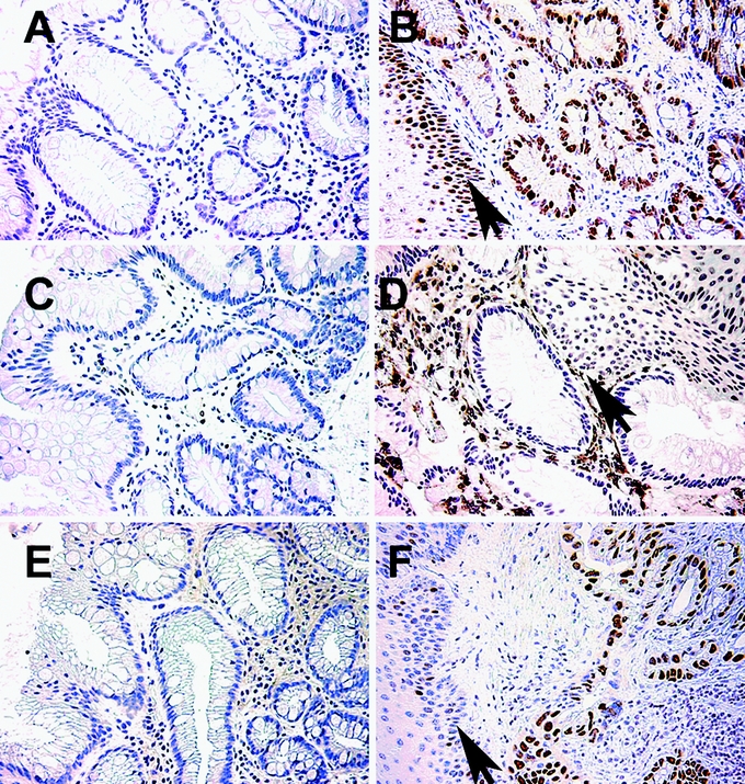
FIGURE 1. Expression patterns of Ki-67, COX-2, and p53 in BE patients prior and after partial ablation. Immunohistologic staining for Ki-67 (A, B), COX-2 (C, D), and p53 (E, F) of BE patient biopsies prior (A, C, E) and after MPEC ablation (B, D, F). Note the intense brown staining of tissues in the biopsies that were taken after ablation. Arrows indicate squamous epithelium.