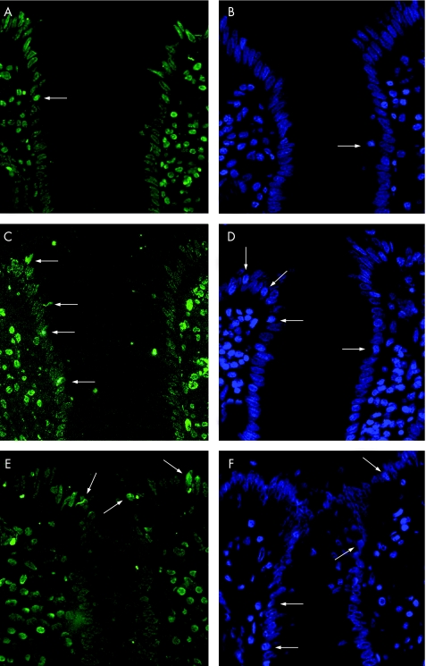
Figure 7 Terminal deoxynucleotidyl transferase‐mediated deoxyuridine triphosphate nick‐end labelling (A,C,E) and 4′,6‐diamidino‐2‐phenylindole (B,D,F) stained thin sections of sigmoid colon from control (A,B), active Crohn's disease (C,D) and active ulcerative colitis (E,F). In all pictures, the entrance of a crypt surrounded by surface epithelium is shown. Arrows indicate apoptotic epithelial cells which were much more common in active Crohn's disease and ulcerative colitis when compared with controls. Magnification ×200.

An official website of the United States government
Here's how you know
Official websites use .gov
A
.gov website belongs to an official
government organization in the United States.
Secure .gov websites use HTTPS
A lock (
) or https:// means you've safely
connected to the .gov website. Share sensitive
information only on official, secure websites.
