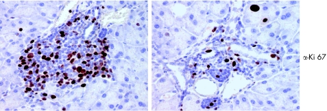
Figure 8 Proliferation of immune cells in the pancreas. Anti‐Ki67 staining of paraffin embedded tissue sections by immunohistochemistry, as described in materials and methods. Positive signals indicate cellular proliferation. Note that a significant number of infiltrating cells show the proliferation marker Ki67.

An official website of the United States government
Here's how you know
Official websites use .gov
A
.gov website belongs to an official
government organization in the United States.
Secure .gov websites use HTTPS
A lock (
) or https:// means you've safely
connected to the .gov website. Share sensitive
information only on official, secure websites.
