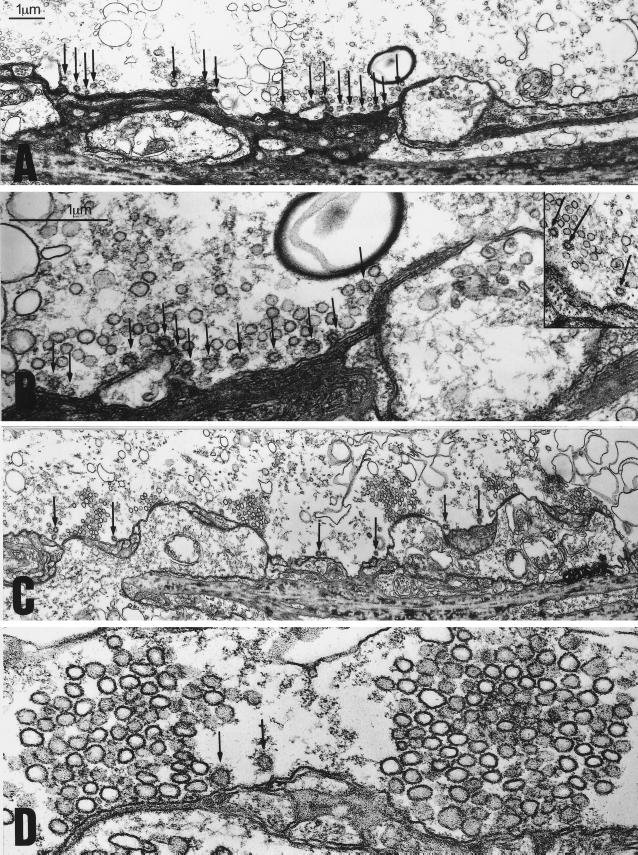
Figure 4.
Injection of GST–SYP into the squid giant synapse preterminal results in a depletion of the vesicle pool and an increase in the number of CCVs during high-frequency stimulation. (A and B) Electronmicrographs from cross sections of a GST–SYP injected synapse. (A) A low magnification image showing several active zones with very small SV clusters. Numerous CCVs are indicated by the arrows. (B) A higher magnification image of the same synapse showing an active zone with a small number of SVs and numerous CCVs (arrows) at some distance from the active zone. Note the numerous uncoated vesicles directly behind the CCVs at the membrane. The Inset in B shows clear examples of CCVs which had pinched off from the plasma membrane in GST–SYP treated synapses. (C and D) Electronmicrographs from cross sections of a GST-injected synapse. (C) A low magnification image showing several active zones with much larger vesicle clusters. A few CCVs (arrows) are seen between the active zones. (D) A higher magnification image showing two active zones with their typical cluster of SVs.

