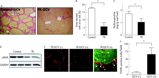
Figure 7 Effect of 17COL promoter‐induced thymidine kinase on the progression of thioacetamide (TAA)‐induced hepatic fibrosis. Rats were treated as shown in fig 1B, iii. (A) Sirius red staining. Fibrotic septum caused by TAA treatment (control) was dramatically suppressed by ganciclovir (GCV) treatment together with the expression of thymidine kinase under the control of 17COL promoter (thymidine kinase‐GCV). LNL‐GFP was coinfected as control. (B, C) Estimation of liver fibrosis. The degree of hepatic fibrosis was quantified by measuring the area positive for Sirius red‐staining (B) and hydroxyproline content (C). Data obtained from six rats in each group are the mean (SD). *p<0.05. (D) Western blot. Lysates of the livers were prepared from two groups (control‐GCV and thymidine kinase‐GCV). Expression of smooth muscle‐α actin (αSMA) and glyceraldehyde‐3‐phosphate dehydrogenase (GAPDH) was determined by immunoblotting. (E) Double immunofluorescence staining for αSMA (red) and TUNEL (green). TUNEL‐positive cells were few in number in the liver infected with 17COL‐thymidine kinase (left). After GCV injection, TUNEL‐positive cells became apparent along αSMA‐positive fibrotic septae (middle). Higher magnification indicates that TUNEL positivity was observed in αSMA‐positive cells in fibrotic septae (arrowheads) and was also seen in αSMA‐positive parenchymal sinusoidal HSCs (arrows; right). (F) Estimation of TUNEL‐positive cells. For semiquantitative analysis of cell death, the number of TUNEL‐positive cell per field was counted in 10 fields from each slide randomly selected. TUNEL‐positive cell numbers increased 10‐fold in GCV‐injected rat livers compared to levels in non‐treated rats. GCV. *P<0.05

An official website of the United States government
Here's how you know
Official websites use .gov
A
.gov website belongs to an official
government organization in the United States.
Secure .gov websites use HTTPS
A lock (
) or https:// means you've safely
connected to the .gov website. Share sensitive
information only on official, secure websites.
