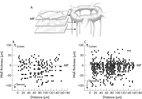
Figure 3 Topography of the 2,4,6‐trinitrobenzenesulphonic acid (TNBS)‐induced mononuclear infiltrate within the colon wall 4 days after the instillation of antigen. Serial optical sections of a 1‐mm2 grid were analysed in whole mounts of the colon wall for the presence of infiltrating mononuclear cells. The linearly encoded optical sections (5 μm intervals) were obtained for 400 μm thickness of the colon wall. In each section, the infiltrating mononuclear cells were counted using the MetaMorph cell counting application threshold on cell nuclei. The nuclei of crypt epithelial cells, readily identifiable by their distinctive structural orientation, were excluded from the analysis. The results of representative (A) control (n = 25) and (B) TNBS‐treated (n = 25) mice are shown. The mucosal capillary plexus was arbitrarily defined as 0 (line) with positive numbers extending to the lumenal surface and negative numbers extending to the serosal surface. MP, mucosal plexus.

An official website of the United States government
Here's how you know
Official websites use .gov
A
.gov website belongs to an official
government organization in the United States.
Secure .gov websites use HTTPS
A lock (
) or https:// means you've safely
connected to the .gov website. Share sensitive
information only on official, secure websites.
