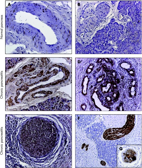
Figure 2 Immunohistochemical analysis of artemin in normal pancreas (A, B) and in chronic pancreatitis (D–G). In normal pancreatic tissue samples, immunostaining of artemin was only faintly present in the smooth muscle cells of arteries (see arrows in (A)). Acini, and intrapancreatic nerves and ducts did not reveal artemin immunoreactivity. In chronic pancreatitis, artemin showed increased immunoreactivity in the smooth muscle cells of arteries (C), in tubular complexes (D) and in the neural components (E–G). In nerves, artemin was present in the cytoplasm and nuclei of Schwann cells (E, F) and in intrapancreatic neural ganglia (G). Magnification 200× in all images except E; E 100×; chromogen: DAB.

An official website of the United States government
Here's how you know
Official websites use .gov
A
.gov website belongs to an official
government organization in the United States.
Secure .gov websites use HTTPS
A lock (
) or https:// means you've safely
connected to the .gov website. Share sensitive
information only on official, secure websites.
