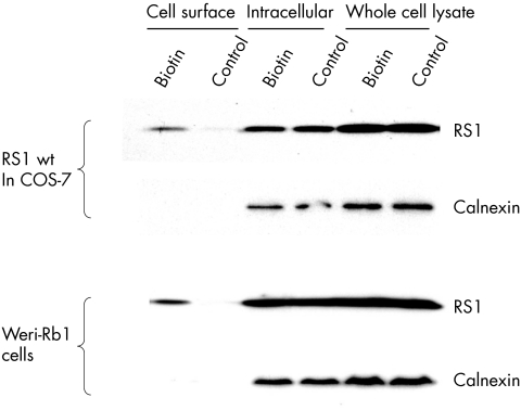
Figure 3 Biotinylation of cell surface proteins. Following biotinylation cells were lysed and fractionated. Control cells were not exposed to biotin but otherwise followed the same protocol. Western blot analysis was used to detect the presence of retinoschisin, or the intracellular control protein (calnexin). Retinoschisin was detected in all fractions including the cell surface in the biotinylated cells but not on the cell surface of the control cells. Calnexin was not detected in the cell surface fractions.

An official website of the United States government
Here's how you know
Official websites use .gov
A
.gov website belongs to an official
government organization in the United States.
Secure .gov websites use HTTPS
A lock (
) or https:// means you've safely
connected to the .gov website. Share sensitive
information only on official, secure websites.
