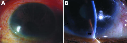
Figure 1 Slit lamp microscopic pictures of ICE patients. (A) Shows iris atrophy, iris holes, and corectopia of a patient with progressive iris atrophy syndrome (patient 1). (B) Shows corneal oedema, iris atrophy (thin arrow), and peripheral anterior synechia (thick arrow) of a patient with Chandler's syndrome (patient 11).

An official website of the United States government
Here's how you know
Official websites use .gov
A
.gov website belongs to an official
government organization in the United States.
Secure .gov websites use HTTPS
A lock (
) or https:// means you've safely
connected to the .gov website. Share sensitive
information only on official, secure websites.
