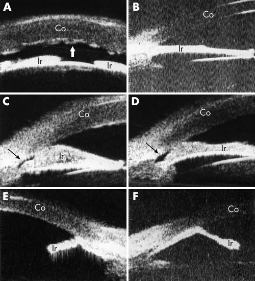
Figure 2 Ultrasound biomicroscopic (UBM) features of ICE syndrome. (A) Shows the shallow anterior chamber and severe corneal oedema in a patient with Chandler's syndrome (patient 5). Folds of Descemet's membrane present a wave‐like appearance (indicated by the white thick arrow). (B) Demonstrates the UBM image of a patient with progressive iris atrophy (patient 2). This patient had extensive iris atrophy and peripheral anterior synechiae, which gave rise to severe glaucoma with IOP of 47 mm Hg and optic nerve atrophy (cup to disc ratio of 0.9). A “bridge‐shaped” peripheral anterior synechia was found in two patients with Chandler's syndrome (patients 10 and 6, respectively), as illustrated in (C) and (D). The iridocorneal angle was indicated by the thin black arrow. Both patients had a shallow anterior chamber with a slightly elevated (patient 10) or normal (patient 6) IOP. (E) and (F) demonstrate the iridocorneal angle with an “arborised” shape caused by severe iris atrophy and broad based peripheral anterior synechiae in two patients with Cogan‐Reese syndrome (patients 15 and 16). Both patients had severe glaucoma as judged by IOP elevation and optic nerve cupping. Co, cornea; Ir, iris.

An official website of the United States government
Here's how you know
Official websites use .gov
A
.gov website belongs to an official
government organization in the United States.
Secure .gov websites use HTTPS
A lock (
) or https:// means you've safely
connected to the .gov website. Share sensitive
information only on official, secure websites.
