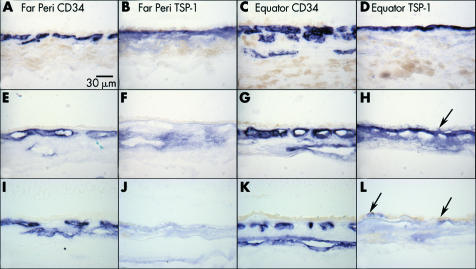
Figure 2 Distribution of TSP‐1 in the choroid. TSP‐1 expression in the choroid from a 3 year old eye (A–D; case 1), a normal 70 year old eye (E–H; case 2), and an 81 year old eye with early AMD (I–L; case 19). Immunoreactivity for TSP‐1 is intense at the equator and faint at ora serrata in Bruch's membrane of both young (B, D) and aged control (F, H) eyes. No dramatic change in TSP‐1 staining is observed between young and old eyes at the equator. In the early AMD eye, no remarkable TSP‐1 staining is observed in the far periphery (J) or at the equator (L). Negative to faint TSP‐1 staining is observed in RPE both in aged control eyes (H, arrow) and eyes with early AMD (L, arrow). CD 34 staining shows choriocapillaris and large choroidal blood vessels (A, C, E, G).

An official website of the United States government
Here's how you know
Official websites use .gov
A
.gov website belongs to an official
government organization in the United States.
Secure .gov websites use HTTPS
A lock (
) or https:// means you've safely
connected to the .gov website. Share sensitive
information only on official, secure websites.
