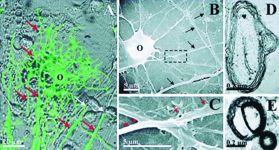
Figure 3.
(A) ES cell-derived oligodendrocytes are capable of myelinating multiple axons in culture. O1 immunoreactivity (green) is superimposed on a phase-contrast image in a mixed ES cell-derived neuronal/glial culture (9 DIV). White arrows indicate axons and red arrows indicate O1 immunoreactive wrapped axon segments. (B) Scanning EM shows oligodendrocyte (O) and passing axons (black arrows). Higher magnification (box from B) demonstrates early axon wrapping (red arrows) by an oligodendrocyte process (C), which is similar to early phases of myelination described in studies using video microscopy (49). Transmission EM shows myelin profiles typical of early myelination in 9 DIV culture (D and E).

