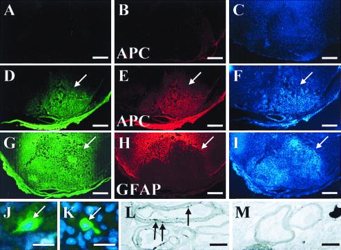
Figure 5.
Transplanted 4−/4+ stage EB cells are capable of differentiating into myelin forming oligodendrocytes in demyelinated adult rat spinal cord. Partial coronal cross-sections (dorsal surface downward) from rats that received sham-transplantation (A–C) or ES cell transplantation (D–F and G–I, two separate animals) demonstrate immunoreactivity for anti-M2 (A, D, and G), anti-APC (B and E) and anti-GFAP (H). Anti-M2 is a mouse-specific Ab for recognition of transplanted mouse ES cells (37), whereas anti-APC labels oligodendrocytes (40). Increased nuclear density at the site of transplantation is indicated by corresponding Hoechst 33342 staining (arrows; F and I) compared with control (C). ES cell-derived oligodendrocytes primarily occupy the zone of demyelination, whereas there is a corresponding paucity of GFAP immunoreactivity in this area (arrows; D, E, G, and H). High magnification shows a native MBP immunoreactive cell (green) from the ventral column distant from the site of transplantation (white arrow, J) and a probable transplanted MBP immunoreactive cell from the center of the area of transplant (white arrow, K). Transmission EM shows early M2-positive myelin (black arrows indicate DAB precipitate particles associated with M2 immunoreactivity) produced by a transplanted ES cell-derived oligodendrocytes (L), and early M2-negative native myelin (M) in ultrathin sections without EM staining. Typical EM myelin contrast staining was not done to be able to visualize the diaminobenzidine (DAB) precipitate associated with anti-M2 immunoreactivity. Scale bars: A–I = 1 mm; J and K = 20 μm; L and M = 0.5 μm.

