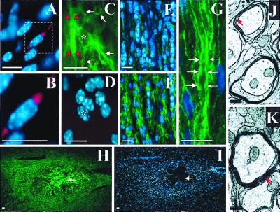
Figure 6.
ES oligosphere-derived cells can migrate and myelinate axons when transplanted into dysmyelinated spinal cords of adult shiverer mice, which lack the gene to produce MBP (27–31). Transplanted cells were identified by Cell Tracker Orange epifluorescence (red) or immunoreactivity for MBP (green). Hoechst 33342 (blue). Cell Tracker Orange-labeled cells were found to align with native intrafascicular oligodendrocytes in white matter (A and B). An ES cell-derived (MBP immunoreactive) oligodendrocyte (asterisk) with longitudinally oriented processes (white arrows) is shown in C and D. Red arrows mark probable myelination around an adjacent axon (C). Little MBP immunoreactivity is present in white matter of a longitudinal spinal cord section from a mouse that received sham transplantation (E). A gradient of MBP immunoreactivity is centered on the site of ES cell transplantation (F). (G) High magnification shows intrafascicular oligodendrocyte nuclei (blue) and MBP immunoreactivity (green) characteristic of axonal myelination (white arrows; refs. 43 and 44) in white matter from a mouse that received ES cell transplantation. The spatial distribution of MBP immunoreactivity, 1 month after ES cell transplantation, is shown at low magnification (H) with corresponding Hoechst 33342 counterstaining (I). White arrows indicate the center of the transplant. Transmission EM shows four loose wraps of myelin, which represents the maximal number of layers typically seen around axons in control animals (red arrow, J), and 9 or greater compact wraps around axons from the area of the transplant (red arrow, K). shiverer mutant mice lack a functional MBP gene that is required to form mature compact myelin; therefore, the presence of mature compact myelin is a gold standard for transplant oligodendrocyte associated myelin. Scale bars: A–I = 10 μm; J and K = 0.3 μm.

