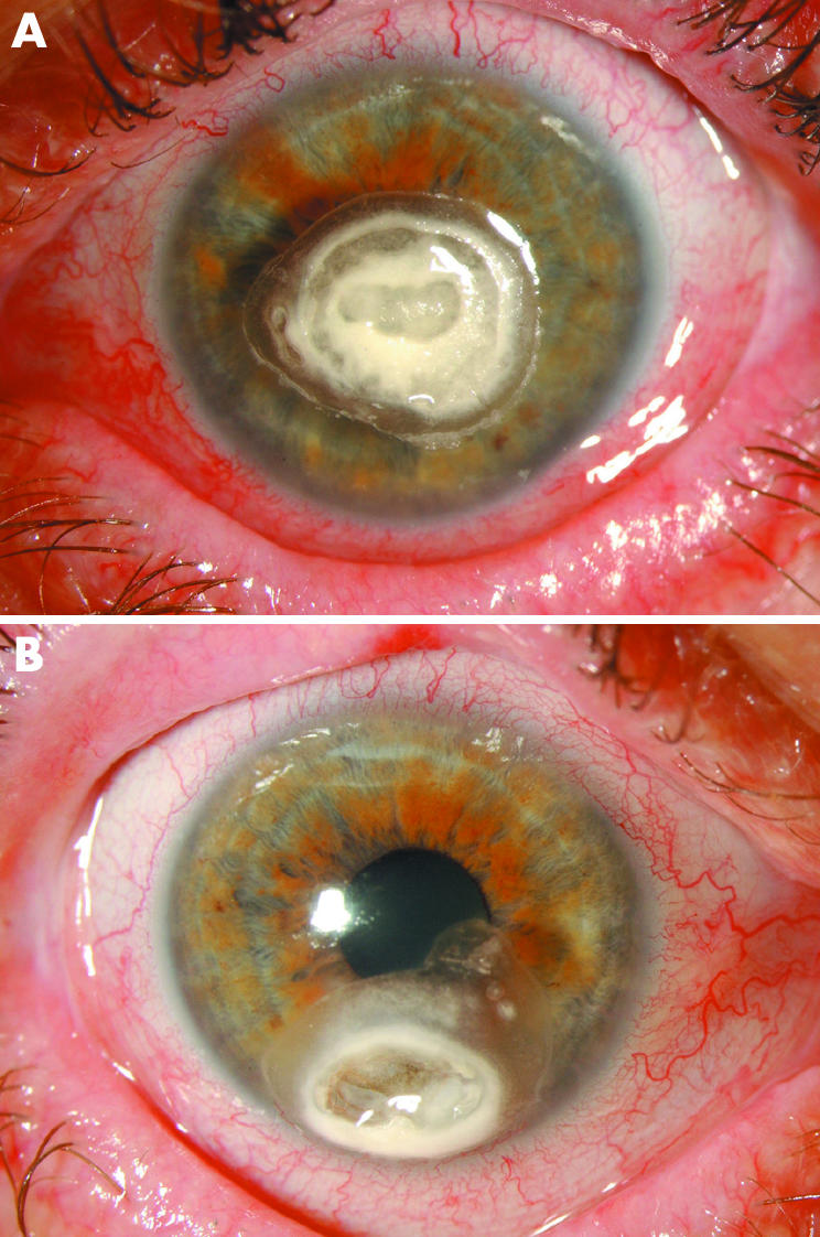
Figure 1 (A) Case 1 left eye. The grey white calcium phosphate deposits recurred within days after an attempt to remove them by superficial keratectomy. The calcification extended into the deep stroma. (B) Case 1 right eye. Calcified plaque that developed within 48 hours in an area previously affected by epithelial keratopathy following trichiasis. Note corneal thinning within the plaque that developed over 2 months and required emergency repair.
