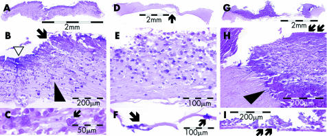
Figure 3 Case 1, light microscopy. The low power photomicrographs (A), (D), and (G) show the extent of the calcifications in both the left and the right cornea, affecting the entire thickness of the specimens. The calcifications are slightly more purple than the non‐calcified corneal stroma in these haematoxylin and eosin stained sections of cornea. The higher power photomicrographs (B), (E), and (H) show the variable histological appearance of the calcifications. The calcifications could consist of small concentrically laminated spherules (B) and (E), they could be confluent (indicated by the arrow in (B)), and form areas of solid plaque (arrowhead in (H)). Solid plaque formation could be seen in the centre of the lesion (open arrowhead in (B)) or at the periphery (arrowhead in (H)). The margin of affected corneal stroma could be sharp (arrowhead in (H)) or consist of a sprinkling of individually calcified spherules (arrowhead in (B)). Descemet's membrane of the left eye was detached from the corneal stroma after histotechnical processing (indicated by an arrowhead in (D)). It is therefore represented in the separate photomicrograph (F). Bowman's membrane (arrows in (H)) and Descemet's membrane (arrows in (F) and (I)) were found to show calcifications in both eyes.

An official website of the United States government
Here's how you know
Official websites use .gov
A
.gov website belongs to an official
government organization in the United States.
Secure .gov websites use HTTPS
A lock (
) or https:// means you've safely
connected to the .gov website. Share sensitive
information only on official, secure websites.
