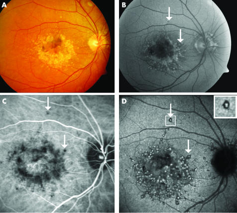
Figure 1 Autofluorescent and indocyanine green angiography features of drusen in malattia leventinese. Case 2, right eye. (A) Colour fundus photograph shows large round drusen aggregated in the paracentral area and small radial drusen located temporally to the macula. (B) Large drusen, in the macular area and around the optic disc, are clearly autofluorescent, whereas small radial drusen are hardly detectable. An appearance of central sparing is observed. (C) In the early phase of the ICG sequence (2 minutes) several dots of hypofluorescence are distributed around the macular area approximating the location of the large round drusen. White arrows indicate some isolated large drusen. (D) In the late phase (30th minute), the large round drusen appear as hyperfluorescent spots surrounded by hypofluorescent halos (white arrows). The small radial drusen cannot be clearly seen in any of the ICG frames.

An official website of the United States government
Here's how you know
Official websites use .gov
A
.gov website belongs to an official
government organization in the United States.
Secure .gov websites use HTTPS
A lock (
) or https:// means you've safely
connected to the .gov website. Share sensitive
information only on official, secure websites.
