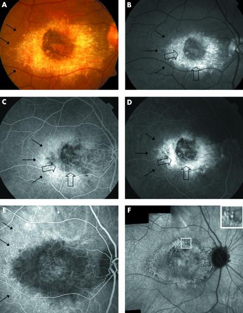
Figure 3 Fluorescein angiography (FA) compared to indocyanine green features in malattia leventinese. Case 4, right eye. (A) Colour fundus photograph shows large drusen aggregated around the macular area. Small radial drusen are indicated by thin black arrows. (B) On the red free frame, the large drusen (indicated by large open arrows) are located around the macular area with a relative sparing of the centre. The large round drusen appear brighter than the small radial ones. The small radial peripheral drusen are in dotted lines (thin black arrows). (C) In the early phase of the FA sequence (30 seconds) we observe, from the centre of the macula to the periphery, a dark area corresponding to the relative central sparing, a grey zone harbouring a buoy pattern (open arrows), surrounded by an ill defined zone of hyperfluorescence and the intense staining of the small radial drusen (small black arrows). (D) At 4 minutes 30 seconds of the FA sequence, the centre of the macular area remains dark and is surrounded by an ill defined zone of hyperfluorescence progressively stained by the dye (open black arrows). The large round drusen are fuzzy and not clearly discernible. The hyperfluorescence of the small radial drusen is still visible although less intense (small black arrows). (E) In the early phase (2 minutes) of the ICG sequence an ovoid hypofluorescent zone surrounds the central macular area. On the periphery of this zone, the small radial drusen appear hyperfluorescent (black arrows). (F) In the 30th minute of the ICG, the composite frame shows aggregation of the hyperfluorescent large drusen which are distributed around the macular area. Most of the hypofluorescent halos are confluent suggesting a confluence of the large drusen. The temporal small drusen are not visible any more. Square shows an enlarged view of the hyperfluorescent dots surrounded by halos of hypofluorescence.

An official website of the United States government
Here's how you know
Official websites use .gov
A
.gov website belongs to an official
government organization in the United States.
Secure .gov websites use HTTPS
A lock (
) or https:// means you've safely
connected to the .gov website. Share sensitive
information only on official, secure websites.
