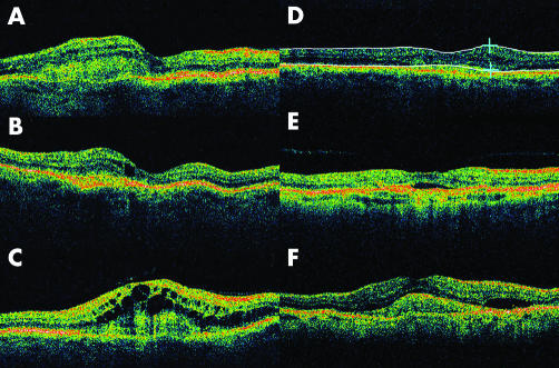
Figure 1 Illustration of OCT findings. The subclassification of intraretinal fluid: (A) sponge‐like retinal appearance with the presence of intraretinal cysts; (B) a single foveal cyst; (C) gross cystoid macular oedema; (D) callipers used to measure retinal thickness, in this case 350 μm; (E) subretinal fluid (SRF) and vitreomacular traction; (F) choroidal neovascularisation complex, SRF, and intraretinal thickening.

An official website of the United States government
Here's how you know
Official websites use .gov
A
.gov website belongs to an official
government organization in the United States.
Secure .gov websites use HTTPS
A lock (
) or https:// means you've safely
connected to the .gov website. Share sensitive
information only on official, secure websites.
