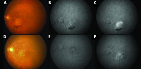
Figure 2 (A) Pretreatment fundus photograph of a patient with subfoveal CNV and haemorrhage at the inferior border. The patient's best corrected visual acuity was 20/200. (B) Early phase of fluorescein angiogram demonstrated a predominately classic subfoveal CNV with (C) leakage in the late phase. (D) Fundus photograph 3 months after a single session of combined PDT with IVTA showing resolution of the haemorrhage with fibrosis of the CNV. The patient's vision improved to 20/100. (E) Early and (F) late phases of fluorescein angiogram showed resolution of the CNV with no evidence of leakage.

An official website of the United States government
Here's how you know
Official websites use .gov
A
.gov website belongs to an official
government organization in the United States.
Secure .gov websites use HTTPS
A lock (
) or https:// means you've safely
connected to the .gov website. Share sensitive
information only on official, secure websites.
