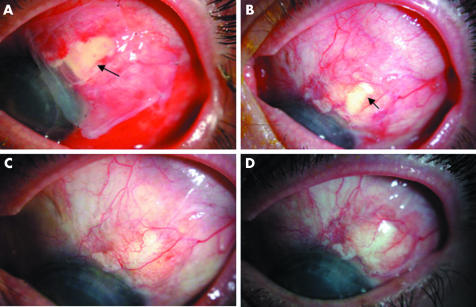
Figure 2 Chronological re‐epithelialisation of epithelial defect following conjunctival erosin repair. (A) Amniotic membrane (outer, epithelial surface down) covering epithelial defect (arrow) day 1 following surgery. (B) Remaining epithelial defect at 2 weeks (arrow) migrating over inner amniotic membrane remnant, outer amniotic membrane has come off. (C) Re‐epithelialisation complete at 4 weeks. (D) Stable re‐epithelialisation at 6 months following surgery.

An official website of the United States government
Here's how you know
Official websites use .gov
A
.gov website belongs to an official
government organization in the United States.
Secure .gov websites use HTTPS
A lock (
) or https:// means you've safely
connected to the .gov website. Share sensitive
information only on official, secure websites.
