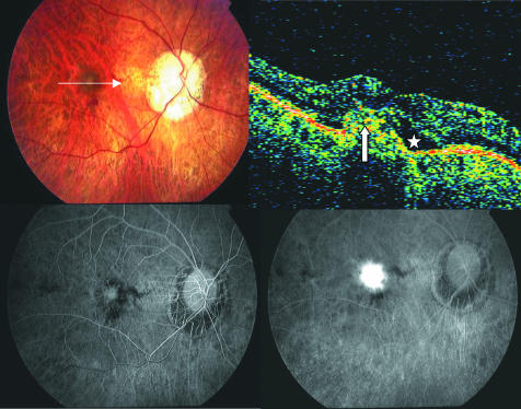
Figure 1 Pretreatment of the right eye of a patient with pathological myopia. Top left, fundus photograph shows characteristic of pathological myopia and signs of choroidal neovascularisation (CNV). Top right, optical coherence tomography (OCT) shows subretinal fluid (asterisk). The CNV complex also is identifiable in OCT scan (white arrow). Bottom left, early phase of angiogram shows a classic CNV membrane. Bottom right, leakage on a late phase angiogram.

An official website of the United States government
Here's how you know
Official websites use .gov
A
.gov website belongs to an official
government organization in the United States.
Secure .gov websites use HTTPS
A lock (
) or https:// means you've safely
connected to the .gov website. Share sensitive
information only on official, secure websites.
