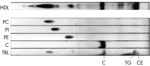
Figure 4 TLC separation and identification of some lipids bound to HDL. Following incubation with RPE cells fed 14C labelled POS, HDL bound lipids were extracted and separated by TLC (bottom of plate at the left). Standards for pure phosphatidyl choline (PC), phosphatidyl inosotol (PI), phosphatidyl ethanolamine (PE), and cholesterol (C) were run, as well as triglyceride rich lipids (TRL) which contains triglycerides (TG) and cholesterol esters (CE) were run.

An official website of the United States government
Here's how you know
Official websites use .gov
A
.gov website belongs to an official
government organization in the United States.
Secure .gov websites use HTTPS
A lock (
) or https:// means you've safely
connected to the .gov website. Share sensitive
information only on official, secure websites.
