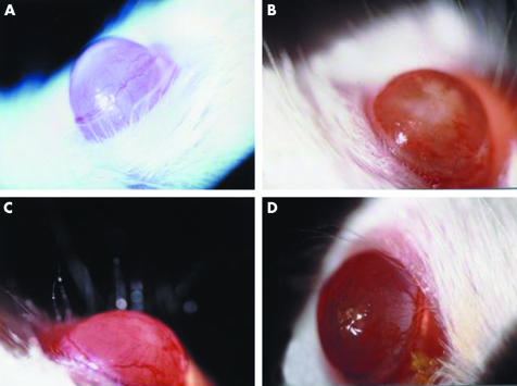
Figure 1 Macroscopic appearance of representative SCID mouse corneas infected with IL‐8/Ad5; 5 μl of IL‐8/Ad5 or LacZ/Ad5 (2.5×10⁷ PFU) was applied to the centre of SCID mouse corneas. (A) Cornea at 12 hours after IL‐8/Ad5 infection. (B, C) Cornea at 16 hours after IL‐8/Ad5 infection. (D) Cornea at 24 hours after IL‐8/Ad5 infection.

An official website of the United States government
Here's how you know
Official websites use .gov
A
.gov website belongs to an official
government organization in the United States.
Secure .gov websites use HTTPS
A lock (
) or https:// means you've safely
connected to the .gov website. Share sensitive
information only on official, secure websites.
