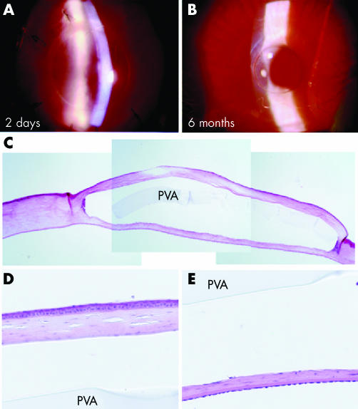
Figure 2 Surgical results of DLKPro. Postoperative slit photographs 2 days (A, ×22) and 6 months (B, ×16) following surgery. HE staining of the same cornea shows an intact disc within the stroma with minimal cellular infiltration (C). High magnification of the flap (D) and posterior stroma (E) shows no cellular infiltration. The space between stroma and implant is an artefact of tissue fixation. PVA = PVA implant.

An official website of the United States government
Here's how you know
Official websites use .gov
A
.gov website belongs to an official
government organization in the United States.
Secure .gov websites use HTTPS
A lock (
) or https:// means you've safely
connected to the .gov website. Share sensitive
information only on official, secure websites.
