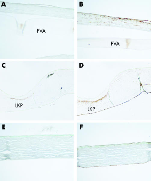
Figure 3 Immunohistochemistry against α‐SMA (A) shows minimal staining compared with the lamellar keratoplasty control (C), indicating the absence of myofibroblasts. Vimentin (B) is expressed locally beneath the epithelium similar to LKP control (D) representing activated fibroblasts. (E, F) Normal control cornea stained with anti‐αSMA (E) and anti‐vimentin (F).

An official website of the United States government
Here's how you know
Official websites use .gov
A
.gov website belongs to an official
government organization in the United States.
Secure .gov websites use HTTPS
A lock (
) or https:// means you've safely
connected to the .gov website. Share sensitive
information only on official, secure websites.
