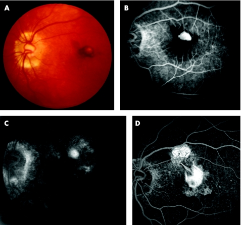
Figure 1 (A) Pre‐photodynamic therapy (PDT) fundus colour photograph showing a juxtafoveal choroidal neovascularisation (CNV) with blood under the foveal centre. (B) Pre‐PDT early phase fluorescein angiography (FA) demonstrates hyperfluorescence of a classic CNV surrounded by blocked fluorescence (subretinal blood). (C) Seven months post‐PDT late phase FA. The CNV is reduced to a smaller staining lesion. Note the RPE changes and absence of leakage from CNV. (D) 26 months post‐PDT late phase FA. Submacular fibrosis developed at the site of previous CNV and in the superior macula.

An official website of the United States government
Here's how you know
Official websites use .gov
A
.gov website belongs to an official
government organization in the United States.
Secure .gov websites use HTTPS
A lock (
) or https:// means you've safely
connected to the .gov website. Share sensitive
information only on official, secure websites.
