
Figure 1. Digestion of collagen I by APCE, rFAP, MMP-1, MMP-2 and MMP-9.
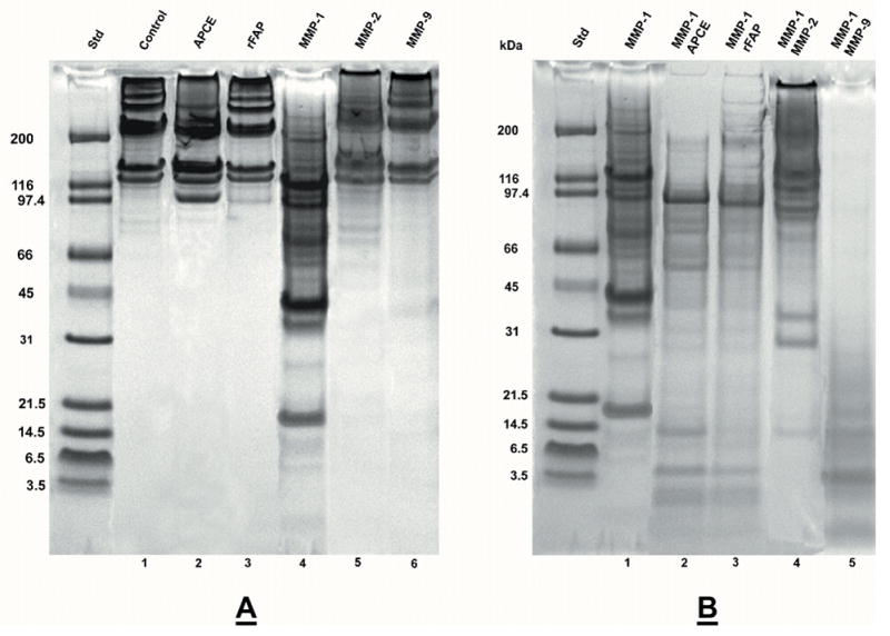
Rat tail collagen type I was gelled at 2 mg/ml and incubated at 37 °C for 24 hours with enzymes in 12.5 mM Na PO4 buffer, pH 7.5. Panel A. Lane: 1) Control-buffer; 2) 16 μg APCE; 3) 16 μg rFAP; 4) 1.5 μg MMP-1; 5) 1 μg MMP-2; 6) 1 μg MMP-9. Panel B. Lane: 1) 1.5 μg MMP-1; 2) 1.5 μg MMP-1, 16 μg APCE; 3) 1.5 μg MMP-1, 16 μg rFAP; 4) 1.5 μg MMP-1, 2.5 μg MMP-2; 5) 1.5 MMP-1, 3.7 μg MMP-9. Digests were reduced and analyzed by 4–12% SDS-PAGE. Where present, the ~97 kDa band is either APCE or rFAP.