
Figure 7.
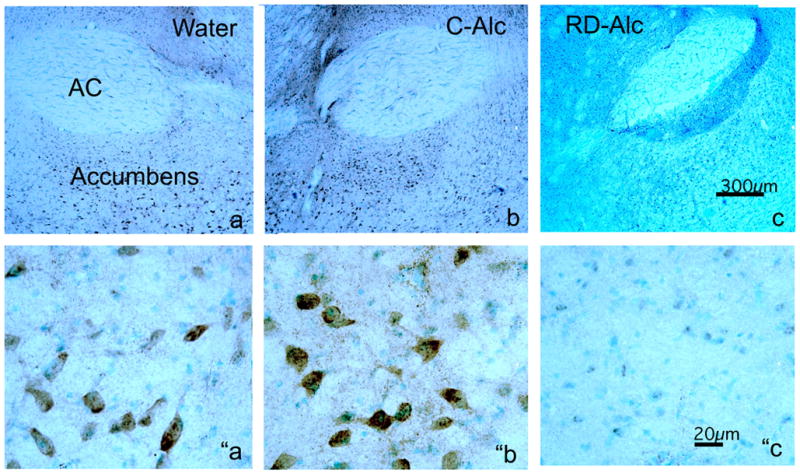
Chronic alcohol on immunostaining (im) of NMDA phosphorylated subunit NR1 (NR1p) in the nucleus accumbens (a,b,c). The intensity of NR1p-im is increased upon continuous chronic alcohol exposure (C-Alc; b and “b) but reduced upon repeated deprivation (RD-Alc; c and “c) as compared with those of the Water control (a and “a). The reduction of the NR1p-im in RD-Alc seem to be more selectively localized in the nucleus accumbens than those shown in Western blot, where entire coronal sections at the nucleus accumbens level were used for analysis. The lower panels are the higher magnification of shell region of nucleus accumbens to show the NR1p-im neurons. AC: anterior commissure, Scale bars: a, b, and c=300μm; “a, ″b, and ”c=20μm.