
Fig. 2.
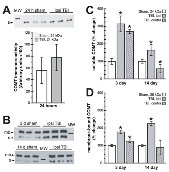
Ipsilateral hippocampal COMT expression levels are chronically altered after TBI. (A) Representative soluble COMT immunoreactivity western blot images detected in 24 h sham and TBI cytosolic extracts, with the summary results shown below. (B) Representative western blot images of total protein extracts from 3 d and 14 d samples detecting both soluble (s, 24 kDa) and membrane-bound (mb, 28 kDa) COMT isoforms. (C) Summary results of changes in ipsi- and contralateral hippocampal soluble COMT immunoreactivity. (D) Summary results of changes in ipsi- and contralateral hippocampal membrane-bound COMT immunoreactivity. Symbols are as follows: open bars-sham; ; shaded, no hatching-ipsilateral; shaded, hatched-contralateral. Marker in (A–B) is 25 kDa. Asterisks (*) indicate p<0.05.