
Fig. 3. Ligation of modified DNA substrates.
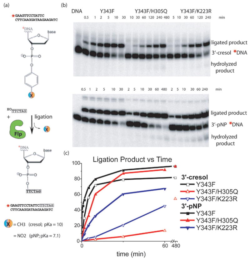
Schematic representation of the DNA ligation assay using a substrate carrying a single Flp binding site with a tyrosine mimic (cresol or pNP) esterified to the 3’ phosphate. A short oligo complementary to the overhang of the bottom strand, which mimics the incoming strand, is added in excess, displaces the tyrosine mimic from the 3’ end of the DNA, and produces a longer ligated product. (b) PAGE separation of ligation products. Note that different timepoints were taken for each Flpe variant. (c) Quantitation of gels showing ligated product as a percentage of total DNA. Reactions catalyzed by all three proteins displayed rate increases with the 3’-pNP compared to the 3’-cresol, when the pKa of the leaving group changed from 10 to 7.1. However, H305Q showed the most dramatic increase.