
Fig. 5.
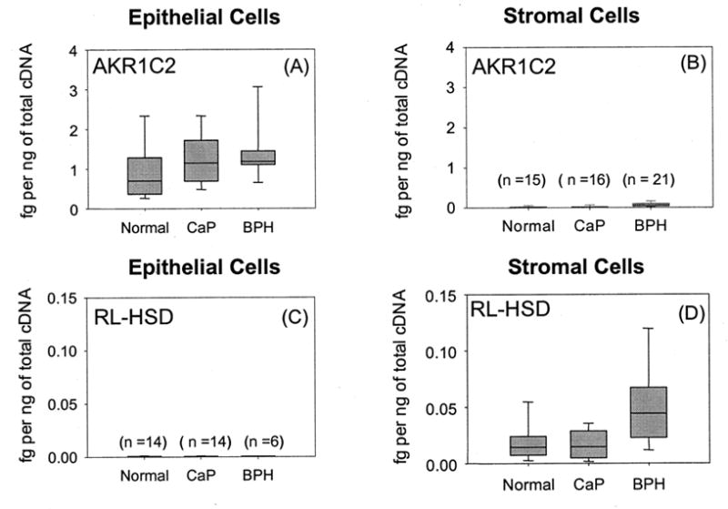
Expression of AKR1C2 and RL-HSD in primary cultures of epithelial and stromal cells taken from normal patients, cancer patients (CaP), and patients with benign prostatic hyperplasia (BPH). Cells were obtained from biopsy samples under an IRB approved protocol obtained by Dr. Donna M. Peehl at the University of Stanford. Cells were cultured as described (Bauman et al, 2006b). AKR1C2 and RL-HSD were quantified by real-time RT-PCR using validated primers (Bauman et al., 2006b) and amounts were normalized to two housekeeping genes (GAPDH-high abundance and porphobilinogen deaminase low abundance) and expressed as fg transcript per ng of total cDNA. The box plots show the median values and their associated standard areas. Adapted from Bauman et al. 2006b.