
Fig. 5.
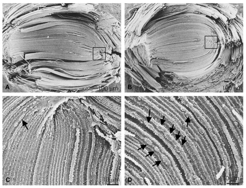
Structural and ultrastructural analysis of lenses overexpressing Cx50. Scanning electron micrographs of non-transgenic (A, C) and transgenic (B, D), line 63 mouse lenses. (A and B) At low magnification, the S-shaped structure of fibers can be seen in both types of lenses. (C and D) At higher magnification, it is clear that the shape and the width of fiber cells in the transgenic lenses are not as uniform as in the non-transgenic lenses and areas with bulges along single fiber cells are evident (arrows). Magnification bars are indicated in each panel.