
Fig. 6.
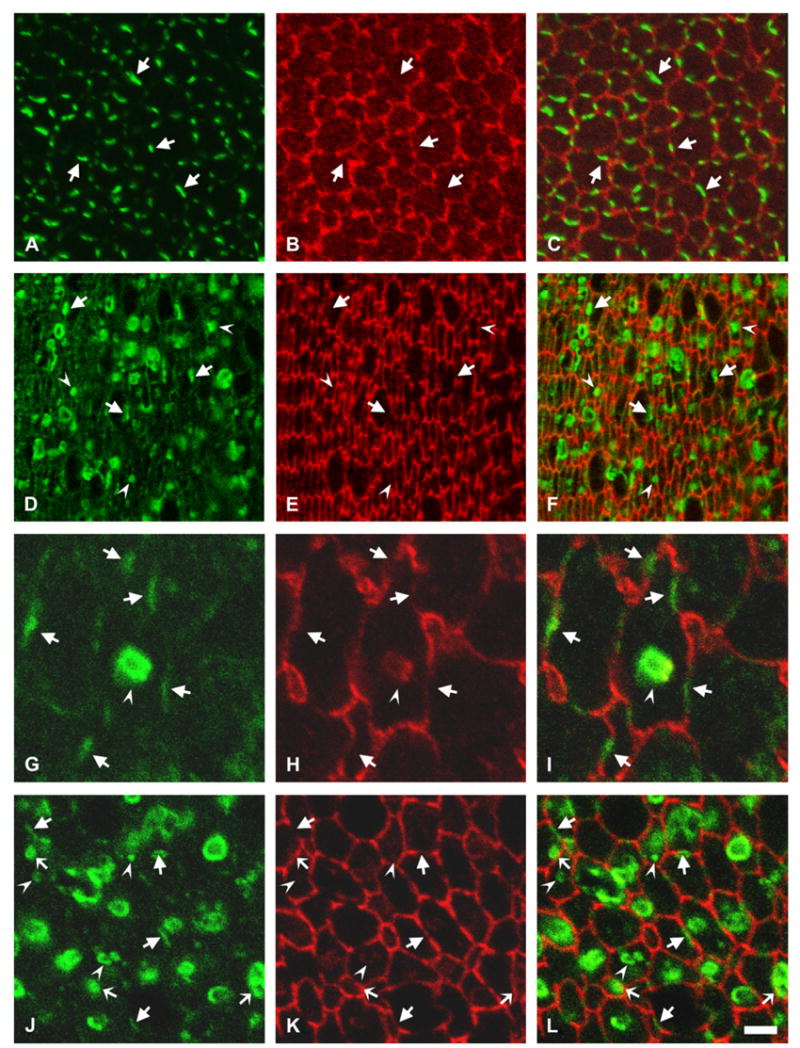
Distribution of the transgene Cx50 protein. (A–C) Photomicrographs show immunolocalization of Cx50 in cross-sections of postnatal day 10 lenses from non-transgenic mice from line 5 after double labeling immunofluorescence using rabbit polyclonal anti-Cx50 antibodies (A) and a mouse monoclonal anti-N-cadherin antibody (B). (D–F) Photomicrographs show immunolocalization of Cx50 in cross-sections of postnatal day 10 lenses from transgenic mice from line 5 after double labeling immunofluorescence using rabbit polyclonal anti-Cx50 antibodies (D) and a mouse monoclonal anti-N-cadherin antibody (E). (G–I) A vesicle-like structure observed at higher magnification after double labeling immunofluorescence with rabbit polyclonal anti-Cx50 antibodies (G) and a mouse monoclonal anti-N-cadherin antibody (H). (J–L) Photomicrographs show immunolocalization of the transgene Cx50 in cross-sections of postnatal day 10 lenses from transgenic mice from line 5 after double labeling immunofluorescence using rabbit polyclonal anti-FLAG antibodies (J) and a mouse monoclonal anti-N-cadherin antibody (K). The immunostaining for Cx50 or the FLAG epitope tag is shown in green and that for N-cadherin is shown in red. The merged images are shown in C, F, I and L. Sections from transgenic mice show abundant immunoreactivity to anti-Cx50 or -FLAG antibodies localized intracellularly (F, L). Irregularities in cell diameter and organization of lens fiber cells are evidenced by the anti-N-cadherin staining (E, K). Anti-Cx50 or anti-FLAG immunoreactivity at the plasma membrane of fiber cells is indicated by arrows; intracellular staining is indicated by arrowheads. Overlap between the anti-FLAG immunoreactivity in vesicles and anti-N-cadherin immunoreactivity at the plasma membrane is indicated by thin arrows in panels J–L. The brightness of the cytoplasmic vesicles in the transgenic lenses precluded the use of the laser beam at the same intensity for image acquisition of lens sections from transgenic and non-transgenic animals; thus, while Cx50 and FLAG immunoreactivities were readily detected along fiber cell membranes in transgenic animals, this is not always apparent in the images shown (compare panels Awith D and J). Bar: 8 μm for A–F, 2 μm for G–I and 4 μm for J–L.