
Fig. 3.
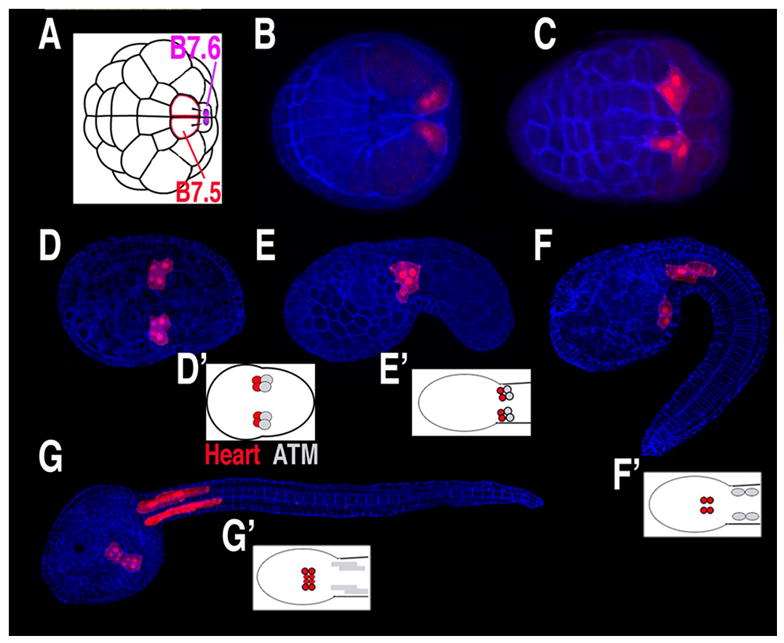
(A–G) Diagrams and confocal micrographs of Ciona embryos, stained with phalloidin (blue) and with B7.5 lineage cells expressing fluorescent proteins under the control of the Mesp enhancer (red), anterior to the left. (A) Diagram of 64-cell stage embryo, position of putative germ plasm in purple. (B) Dorsal view of gastrula stage embryo. (C–D) Early and late neurula stage embryos, ventral view. (E–F) Early and late tailbud stage embryos, lateral view. (G) Larva, ventra-lateral view. (D’–G’) Diagrams of B7.5 lineage fates from a ventral perspective, heart lineage in red, anterior tail muscle (ATM) in grey. (D and G modified from [24]).