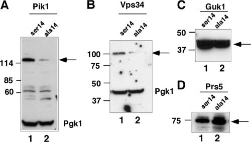
Figure 2.
Analysis of lipid kinases in a cdc37S14A strain. (A) Western blot analysis of TAP-tagged Pik1 (arrow) and Pgk1 in wild-type (ser14) and cdc37S14A (ala14) strains. (B) Analysis of Vps34; details are as described in A. (C and D) Analysis of Guk1 and Prs5 steady-state levels by Western blotting. In each case, the arrow indicates the position of the TAP-tagged protein.

