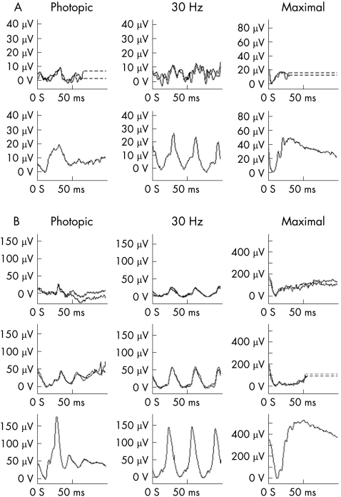
Figure 1 (A) ERGs recorded using a surface electrode on the lower eyelid. The upper trace is from patient 3, the lower trace from a normal child. Note the reduction in the b:a ratio in the photopic single flash ERG; the delayed and reduced 30 Hz flicker ERG; and the profoundly electronegative bright flash dark adapted ERG (maximal). Broken lines have been used to replace blink/eye movement artefact. (B) ERGs recorded using corneal gold foil electrodes. The upper traces are from patient 8; the middle traces are from patient 6 the lower traces are from a normal subject. The ERG characteristics are similar to those described above with additional amplitude changes in the photopic ERG of patient 8. Broken lines have been used to replace blink/eye movement artefact.

An official website of the United States government
Here's how you know
Official websites use .gov
A
.gov website belongs to an official
government organization in the United States.
Secure .gov websites use HTTPS
A lock (
) or https:// means you've safely
connected to the .gov website. Share sensitive
information only on official, secure websites.
