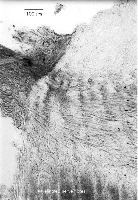
Figure 1 Section of lamina cribrosa illustrating lamina cribrosa thickness (LCT) measurement. LCT was defined as the measurement from the first collagenous plate to the point where myelinated axons became visible, delineated by “X”. The area was arbitrarily divided into four sections (a–d), about 100 μm thick, for measurement of cribrosal beam thickness. Magnification 10×. a, anterior; b, mid‐anterior; c, mid‐posterior; d, posterior.

An official website of the United States government
Here's how you know
Official websites use .gov
A
.gov website belongs to an official
government organization in the United States.
Secure .gov websites use HTTPS
A lock (
) or https:// means you've safely
connected to the .gov website. Share sensitive
information only on official, secure websites.
