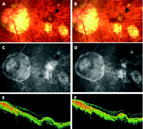
Figure 2 Case 2. (A) Fundus photograph showing the myopic choroidal neovascularisation (mCNV) before treatment (arrowhead). (B) Fundus photograph showing choroidal neovascularisation after treatment (arrowhead). (C) Fluorescein angiography (FA) in the late phase (10 min) showing leakage from the mCNV (arrowhead) before treatment. (D) FA in the late phase (12 min) showing reduced leakage from the mCNV (arrowhead) 4 months after injection. (E) Horizontal optical coherence tomography (OCT) image showing raised retinal reflectivity and a hyper‐reflective lesion under the retina that corresponds to the mCNV before treatment. The thickness of the swollen fovea is 245 µm. (F) OCT image 5 months after intravitreal injection showing the smaller high‐density lesion under the retinal reflectivity compared with that before treatment. The thickness of the foveal retina decreased to 147 µm. The BCVA improved from 0.3 to 0.6.

An official website of the United States government
Here's how you know
Official websites use .gov
A
.gov website belongs to an official
government organization in the United States.
Secure .gov websites use HTTPS
A lock (
) or https:// means you've safely
connected to the .gov website. Share sensitive
information only on official, secure websites.
