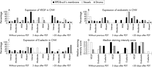
Figure 1 Graphs showing vascular endothelial growth factor (VEGF) (A), endostatin (B) and E‐selectin (C) immunostaining intensity and median staining intensity scores (D) in choroidal neovascular membranes (CNVs) without photodynamic therapy (PDT) and CNV extracted at 3 days and >20 days after PDT. VEGF, endostatin and E‐selectin immunostaining in retinal pigment epithelium (RPE)‐Bruch's membrane, vessels and stromal cells were evaluated separately and semiquantitatively as intense (70–100% positive cells), moderate (40–69% positive cells), mild (1–39% positive cells) or absent. Staining scores of 3, 2, 1 and 0 were assigned to “intense”, “moderate”, “mild” and “absent” intensity of staining, respectively.

An official website of the United States government
Here's how you know
Official websites use .gov
A
.gov website belongs to an official
government organization in the United States.
Secure .gov websites use HTTPS
A lock (
) or https:// means you've safely
connected to the .gov website. Share sensitive
information only on official, secure websites.
