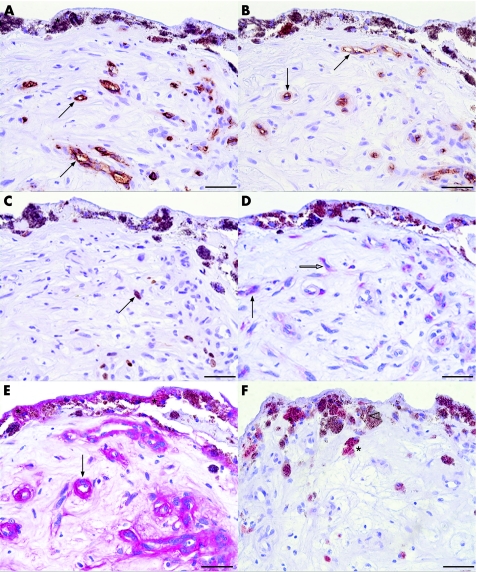
Figure 2 Photomicrographs of surgically excised choroidal neovascular membranes without prior photodynamic therapy. The specimens were probed with antibody against CD34 (A), CD105 (B) and Ki‐67 (C) stained with 3‐diaminobenzidine, resulting in a brown chromogen; vascular endothelial growth factor (VEGF) (D) and endostatin (E) stained with red chromogen; and E‐selectin (F) with 3‐amino‐9‐ethyl carbazole. Haematoxylin was used as a counterstain. CD34 (A) and activated endothelial cell marker CD105 (B) are selectively expressed in vascular structures (arrow). The brown chromogen can be distinguished from the melanin granula (asterisk) contained in pigmented cells. Several cell nuclei express the proliferation marker Ki‐67 (C, arrow). In the serial section of the same specimen (D), VEGF staining was detected within endothelial cells (arrow) and stromal cells (white arrow). In a serial section probed with endostatin, retinal pigment epithelium (RPE)‐Bruch's membrane (asterisks) and vessels (arrow) express endostatin (E). (F) Some RPE cells (asterisk) display E‐selectin immunoreactivity, whereas some RPE cells are not immunoreactive (white arrowhead). Scale bar: 50 μm.

An official website of the United States government
Here's how you know
Official websites use .gov
A
.gov website belongs to an official
government organization in the United States.
Secure .gov websites use HTTPS
A lock (
) or https:// means you've safely
connected to the .gov website. Share sensitive
information only on official, secure websites.
