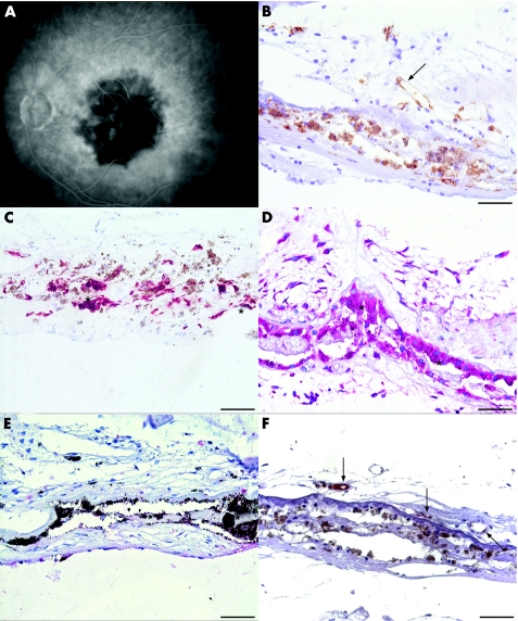
Figure 3 Photomicrographs of choroidal neovascular membranes (CNV) membrane (case 4, table 1) extracted 3 days after photodynamic therapy. Early phase of fluorescein angiography (A) on the day of surgery displays non‐perfusion of the CNV and laser spot area. The serial sections were probed with CD34 (B), cytokeratin 18 (C), vascular endothelial growth factor (VEGF) (D), endostatin (E) and E‐selectin (F). Some vessels depicted by the brown chromogen are patent, but are still lined with damaged endothelial cells (B, arrow). Retinal pigment epithelium (C, asterisks) are strong positive for VEGF (D, asterisks), but not immunoreactive for endostatin (E, asterisk). Endostatin immunoreactivity is absent in CNV (E). Endothelial cells express E‐selectin (F, arrows). Scale bar: 50 μm.

An official website of the United States government
Here's how you know
Official websites use .gov
A
.gov website belongs to an official
government organization in the United States.
Secure .gov websites use HTTPS
A lock (
) or https:// means you've safely
connected to the .gov website. Share sensitive
information only on official, secure websites.
