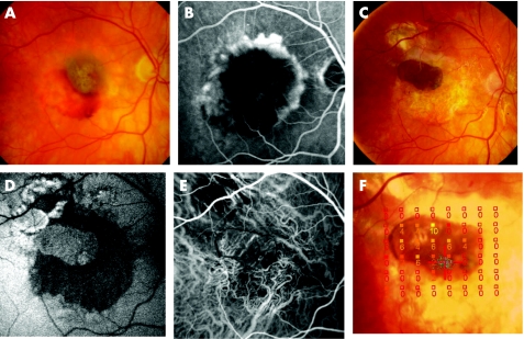
Figure 2 (A–F) Patient 8 presented an occult choroidal neovascularisation with subretinal haemorrhage before operation. Fundus photography (A) and corresponding fluorescein angiography (B); visual acuity = 1.3 logarithm of minimum angel of resolution (logMAR). Six months after surgery, visual acuity improved to 0.16 logMAR. The pigmented retinal pigment epithelium (RPE)–choroid patch is well visible (C). Autofluorescence of the graft was well preserved (D) and indocyanine green angiography shows choroidal vascularisation in or beneath the RPE–choroid sheet (E). Microperimetry showed fixation (blue spots) and retinal sensitivity over the patch area (filled squares, dB light increment sensitivity) (F).

An official website of the United States government
Here's how you know
Official websites use .gov
A
.gov website belongs to an official
government organization in the United States.
Secure .gov websites use HTTPS
A lock (
) or https:// means you've safely
connected to the .gov website. Share sensitive
information only on official, secure websites.
